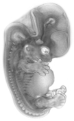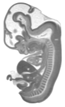
Human Embryo


Human Embryo

Datasets
Datasets Available for Download
(1) Human Embryo MRI's of Carnegie Stages 13-23 (excepting Stage 21)*
(2) Segmented (by organ) Human Embryo MRI's of Carnegie Stages 13-23 (excepting Stage 21)*
These data were produced prior to the establishment of the DICOM standard, therefore, datasets are a series of .tif files.
The .tif image files are 256x256 pixels each.
The datasets are all istropic (the image resolution is equal in x, y, and z dimensions).
Image resolutions range from 39 microns to 156 microns per voxel side (x, y, and z), depending on the age/size of the embryo.
File numbering starts at the caudal end and runs to the cranial end of the embryo.
Within each image slice, dorsal is at the top of the image, ventral at the bottom, anatomical left is at the right of the image, and anatomical right is at the left of the image (i.e., you are looking down toward the caudal direction of the embryo from the cranial end).
More detailed information is included in a readme file with each download, and can also be found on the Technical page of this website.
*These datasets are licensed under a Creative Commons license. Visit the Permissions page of this site for licensing information.